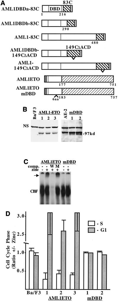
FIG. 9.
ETO slows G1 progression when directed to AML1 target genes. (A) Diagram of AML1-INV fusion proteins, AML1-ETO, and AML1ETOmDBD. The 83 C-terminal INV residues, including the ACD, were linked at position 216, 290, or 480 to AML1B, and 121 C-terminal residues of 149C(ΔACD), lacking the ACD, were linked to AML1B at residue 290 or 480. Six amino acids were inserted between amino acids 145 and 146 in the DNA-binding domain of AML1-ETO to generate AML1ETOmDBD. The N terminus of AML1-ETO differs from that of AML1B. (B) Western blot with an AML1 antiserum, detecting AML1-ETO and AML1-ETOmDBD in Ba/F3 subclones.The location of a nonspecific band, which served as a loading control, is shown (NS). AE-2, AML1-ETO clone 2. (C) Gel shift assay with AML1-ETO and AML1ETOmDBD nuclear extracts was carried out as in Fig. 6D. The arrow indicates the AML1-ETO gel shift complex. (D) The relative proportion of each indicated cell line in the S and G1 cell cycle phases, with and without zinc at 48 h, is shown (mean and standard error of two determinations).

