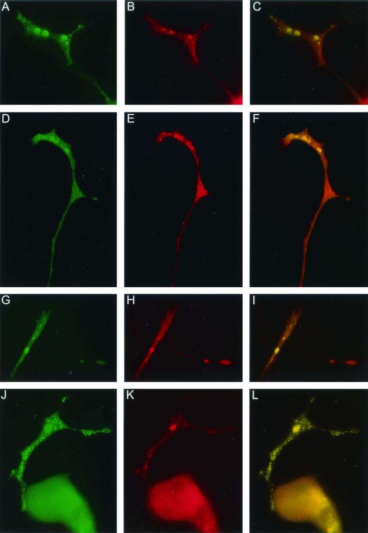
FIG.5.
PC12/FMR7 cells (after 24 h of DOX treatment) double labeled on the one hand for FMRP-GFP (A, D, G, and J) and on the other hand for total RNA (B), ribosomal subunits (P0) (E), kinesin heavy chain (H), and FXR1P (K). For each individual colocalization experiment we have included an image that merges both separate images (green and red). Colocalization results in a yellow color (C, F, I, and L). Photographs show part of a neurite and growth cone with two populations of granules (larger and smaller). Both types of granules show colocalization. Magnifications: A to I, ×2,880; J to L, ×1,600.

