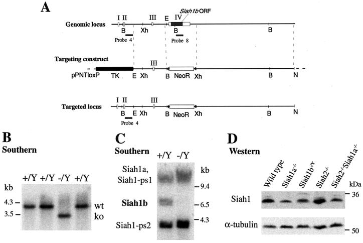
FIG. 1.
Disruption of Siah1b in ES cells by gene targeting. (A) Exons I, II, III, and IV of Siah1b are depicted as open rectangles; the coding region of Siah1b is a filled rectangle. The targeting construct in the vector pPNTloxP is depicted. DNA fragments used as the left and right arms of homology are represented by dotted lines. Homologous recombination yields the targeted locus in which the entire coding region is replaced by a loxP-flanked neomycin resistance gene cassette. B, BamHI; Xh, XhoI; E, EcoRI; triangle, loxP element. (B) Southern analysis of BamHI-digested genomic DNA from wild-type (+/Y) or targeted (−/Y) ES cells, probed with probe 4 (located 5′ of the 5′ targeting arm) to identify the targeted locus. (C) Southern analysis (as for panel B) with coding region probe 8 that hybridizes to the four murine Siah1 genes (including the two pseudogenes Siah1-ps1 and Siah1-ps2), to confirm the loss of the Siah1b gene. (D) Western blot analysis of total protein lysates derived from wild-type, Siah1a−/−, Siah1b−/Y, Siah2−/−, or Siah2−/− Siah1a−/− MEFs, using a monoclonal anti-Siah1 antibody that recognizes both Siah1a and Siah1b.

