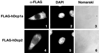
FIG. 5.
Transiently expressed hDcp1a and hDcp2 proteins localize primarily in the cytoplasm. The figure shows the results of immunocytochemical staining of fixed, permeabilized HeLa cells, transiently expressing FLAG-tagged hDcp1a (panels 1 to 3) or hDcp2 (panels 4 to 6). Cells were stained with anti-FLAG M2 monoclonal antibody and visualized with Texas red-conjugated anti-mouse immunoglobulin G antibody (panels 1 and 4). Nuclei are visualized with 4′,6′-diamidino-2-phenylindole (DAPI) (panels 2 and 5). Panels 1 to 6 show one cell, representative of at least 100 observed transfected cells.

