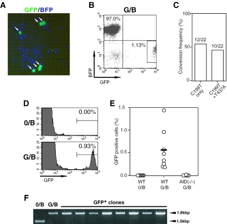
Figure 2.
Gene conversion machinery in DT40 cells can target a non-Ig exogenous gene. (A) Fluorescence microscopic observation of green cells (indicated by arrows) that appeared after 3 weeks culture of G/B construct-bearing DT40 cells, which had initially emitted blue fluorescence exclusively. (B) Loss of the BFP fluorescence in green cells (surrounded by a rectangle) derived from G/B construct-bearing DT40 cells that were initially BFP+GFP− exclusively. Flow cytometric analysis of G/B construct-bearing DT40 cells was carried out after culture for 3 weeks. (C) Mutation profile at the 199th and 437th nucleotides on the BFP gene in G/B construct that was integrated in the IgL locus of DT40 cells. A total of 22 DNA clones of the BFP gene obtained from green cells [shown in (B)] were analyzed. (D) Dependence of green cell generation on the donor GFP gene. Flow cytometric analysis was conducted after culture of 0/B or G/B construct-bearing DT40 cells for 3 weeks. (E) Green cell generation by AID-dependent gene conversion. Wild-type (WT) or AID-deficient DT40 cells were integrated with 0/B or G/B constructs in the IgL locus as indicated. BFP+ clones from each group (n = 8) were analyzed for the appearance of green cells after 3 weeks culture. Data indicate the percentage of GFP+-gated cells [see (D)] in each cultured clone. (F) Retention of structural configuration of the GFP gene in the G/B construct in green cells. The sequence covering the integrated-GFP gene in 9 clones was amplified by PCR using the same primer pair described in Figure 1D. Amplified bands of 1.8 kb were found in all clones tested, suggesting the retention of the GFP genes in the integrated construct. No mutations were found in the GFP gene

