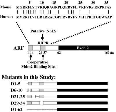
FIG. 1.
Schematic representations of mouse ARF and its mutants. (Top) Alignment of mouse and human ARF protein sequences between amino-terminal residues 1 and 37. Identical amino acids are indicated by bars. (Middle) Structure of the mouse ARF protein, showing the positions of the two reported Mdm2 binding domains (shaded) and the predicted NoLS, RRPR. (Bottom) Structures of mouse ARF deletion mutants analyzed in this study. aa, amino acids.

