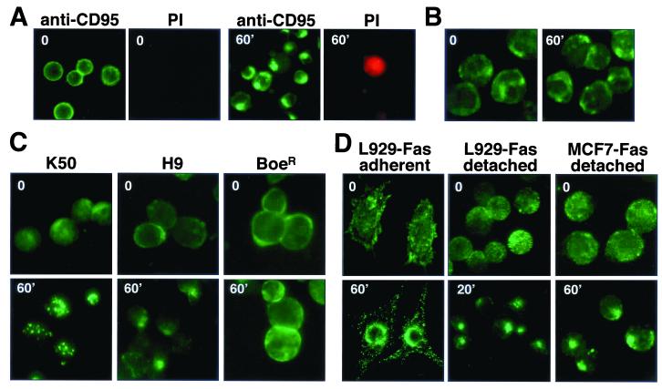
FIG. 2.
Clustering of CD95 is a specific and general effect. (A) Living SKW6.4 cells were treated with FITC-conjugated anti-CD95 and incubated for 60 min on ice (time 0) or for 60 min at 37°C (60′). After stimulation, cells were counterstained with propidium iodide (PI) and analyzed by fluorescence microscopy. (B) To establish specificity of the internalization of CD95, K50 cells were incubated with 1 μg of anti-CD95 per ml for 0 or 60 min and after fixation were stained for CD19 with an FITC-conjugated anti-CD19 mouse MAb. (C and D) CD95 clustering on lymphoid (C) and nonlymphoid (D) cells. Receptor clustering of K50, H9, BoeR, detached L929-Fas, and detached MCF7-Fas cells was performed as follows. Cells were incubated with anti-CD95 followed by FITC-conjugated goat anti-mouse IgG, each for 45 min on ice (t = 0). Cells were then warmed to 37°C and analyzed after the indicated times. After incubation, cells were attached to poly-l-lysine-coated slides and fixed, and samples were analyzed by fluorescence microscopy. Stimulated cells are shown at the same magnification as unstimulated cells. Adherent L929-Fas cells were grown on Polyprep poly-l-lysine slides (Sigma) to 70% confluency and transferred to ice-cold medium. Directly, FITC-conjugated anti-CD95 was added, and cells were incubated for 45 min on ice. After being washed in medium, CD95 clustering was induced by transfer of slides to 37°C medium and incubation for 60 min. After fixation, all cells were analyzed by fluorescence microscopy.

