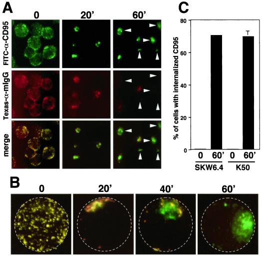
FIG. 3.
Stimulation-dependent internalization of CD95. (A) SKW6.4 cells were incubated with FITC-conjugated anti-CD95 for 45 min on ice followed by incubation at 37°C for the indicated time. After washing, cells were stained with Texas red-conjugated goat anti-mouse IgG (αmIgG) and then transferred onto poly-l-lysine-coated slides, fixed, and analyzed by fluorescence microscopy as described in Materials and Methods. Arrowheads point to cells that did not stain with the secondary antibody and therefore have fully internalized CD95. (B) 2D projections of 3D movies of SKW6.4 cells treated as in panel A. For orientation, the plasma membrane is labeled by a stippled circle. (C) Quantification of CD95 internalization at time point 0 and after stimulation of K50 and SKW6.4 cells for 60 min. The number of cells with 50% or more of CD95 internalized was determined as described in Materials and Methods. The experiment was done in triplicate, and the mean values with standard deviations are shown. The numbers of propidium iodide-positive cells following the incubation at 37°C were 8% for SKW6.4 and 6% for K50, respectively.

