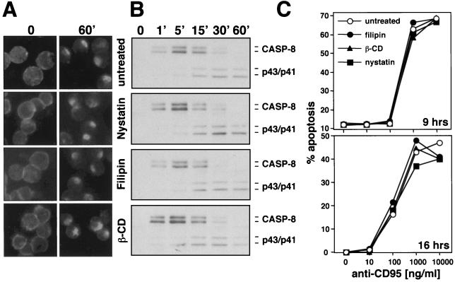
FIG. 7.
CD95 signaling on SKW6.4 cells cannot be inhibited by destruction of lipid rafts (A) SKW6.4 cells were left untreated or were pretreated with 10 μg of nystatin per ml, 1 μg of filipin per ml, or 2 mM methyl-β-cyclodextrin (β-CD) for 1 h at 37°C. Cells were then treated with anti-CD95 and left unstimulated (time 0) or were stimulated for 60 min (60′) at 37°C as described in Fig. 1A. Samples were analyzed by fluorescence microscopy. (B) Analysis of the DISC of 107 SKW6.4 cells treated for different times with the same reagents as in panel A was performed as described in Fig. 1B. (C) Titration of anti-CD95 onto SKW6.4 cells in the presence of the indicated reagents used at the same concentrations as in panel A. Cells were treated in either serum-free medium for 9 h or serum-containing medium for 16 h, and DNA fragmentation was quantified as described in Materials and Methods. Similar data were obtained when H9 cells were tested in the same way (data not shown).

