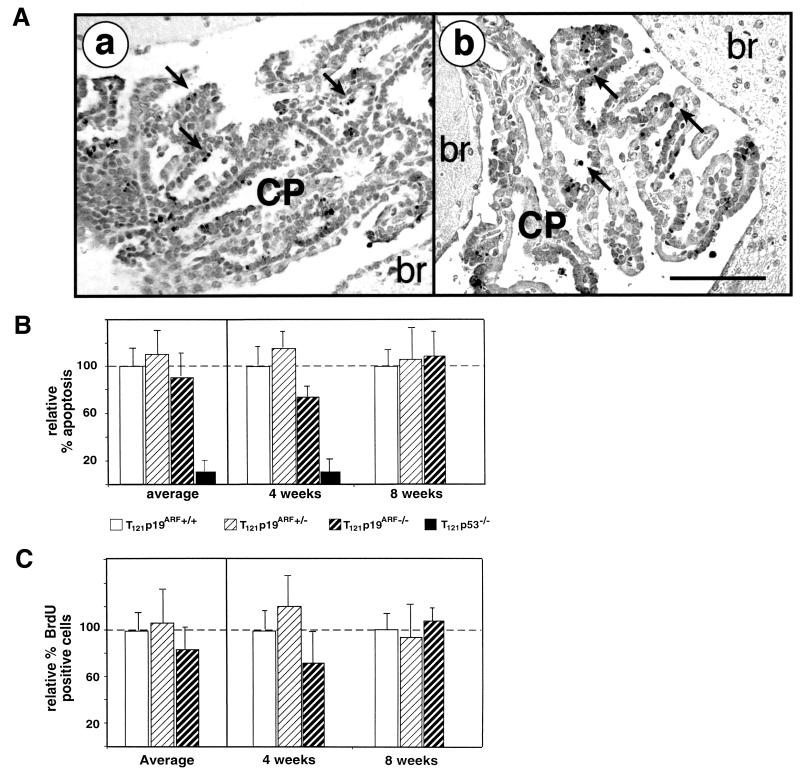
FIG. 3.
T121-induced apoptosis and cell proliferation do not require p19ARF. T121 expression causes abnormal CP cell proliferation and apoptosis. About 85% of the apoptosis is p53-dependent (27) and requires E2F1 (18). The TUNEL assay was used to detect apoptosis (A). Apoptosis levels and morphology were indistinguishable between TgT121;p19ARF−/− (a) and TgT121;p19ARF+/+ (b) CP. Apoptotic nuclei are stained and appear black; representative apoptotic cells are indicated by arrows. The bar (b) is equal to 100 μm; both panels are of the same magnification. Quantitative analysis of apoptosis (B) and proliferation (C) in the CP of three mice each at 4 and 8 weeks of age was carried out. Levels are compared to that of TgT121;p19ARF+/+ tissue, which is considered to represent 100%. Average relative values for all mice of each age and genotype are shown in the left panels. For these data, error bars indicate the variation among mice. In the right panels, data for each mouse are presented, with error bars indicating the field-to-field variation in counts taken from 10 fields per brain. Different mice were used for apoptosis and proliferation assays so as to avoid any impact of BrdU incorporation on apoptosis levels. There is no significant difference in apoptotic indexes between TgT121;p19ARF+/+ and TgT121;p19ARF−/− mice (P = 0.601). Importantly, the index of TgT121;p19ARF−/− CP was significantly higher than that of TgT121;p53−/− CP (P < 0.05). The level of CP cell proliferation was determined by immunostaining of BrdU incorporated in vivo (C). The data show that p19ARF deficiency does not significantly alter cell proliferation (P = 0.288).

