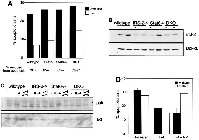
FIG. 5.
IRS-2 and Stat6 are dispensable for the antiapoptotic activities of IL-4. (A) CD4+/CD62L-high T cells were cultured for 24 to 30 h in the presence or absence of IL-4. The percentage of apoptotic cells in the resulting cultures was determined by propidium iodide staining. Percent rescued from apoptosis compares the percentage of apoptotic cells in the IL-4-treated cultures with the percentage of apoptotic cells in untreated cultures and is presented as the average of four independent experiments. *, P < 0.0001 compared to wild type. (B) Enriched T cells were cultured for 17 h in the presence (+) or absence (−) of IL-4. Whole-cell extracts were analyzed for the presence of Bcl-2 by immunoblotting. The blot was stripped and reprobed with an antibody to Bcl-xL. This result is representative of two independent experiments. (C) Enriched and cytokine-starved T cells were cultured in the presence or absence (−) of IL-4 for 3 min. Where indicated, the cells were also preincubated for 10 min with 100 nM wortmannin (wm). Whole-cell extracts were analyzed for the presence of phosphorylated Akt (pakt) by immunoblotting. The blot was stripped and reprobed with an antibody to total cellular Akt. The results are representative of four independent experiments. (D) Lymph node CD4+ T cells were cultured in the presence or absence of 10 ng of IL-4/ml and 100 μg of sodium vanadate/ml (NV) for 24 h. The percentage of apoptotic cells was determined by propidium iodide staining. The results are representative of two independent experiments. The error bars indicate standard deviations.

