
Figure 4.
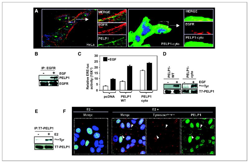
PELP1 interacts with EGFR and promotes EGF-mediated, ligand-independent ER transactivation functions. A, HeLa cells were treated with EGF (50 ng/mL). The cells were then fixed and costained with antibodies against PELP1 (green) and EGFR (red). The images were analyzed by confocal microscopy. Arrows, area of colocalization (yellow) of PELP1 and EGFR (left). PELP1-cyto cells were treated with EGF (50 ng/mL). Cells were stained with antibodies against T7 tag (to detect T7-tagged PELP1-cyto, green) and EGFR (red; right). A small portion of the image showing the colocalization (yellow) was enlarged and shown in the side panels. B, COS-1 cells were serum starved for 24 hours and treated with or without EGF. EGFR was immunoprecipitated by using an EGFR antibody; the presence of PELP1 in the immunoprecipitates was analyzed by Western blotting by using an anti-PELP1 antibody. C, HeLa cells were cotransfected with ER and ERE-luc reporter gene along with pcDNA or PELP1-WT or PELP1-cyto. After 48 hours, cells were stimulated with EGF for 12 hours and ERE-luc reporter activity was measured. D, COS-1 cells were transfected with T7-tagged PELP1-WT and after 72 hours, treated with or without EGF. Total lysates were immunoprecipitated with T7-epitope specific antibody and blotted with phosphotyrosine and T7 antibodies (left). MCF-7 cells that stably express T7-tagged PELP1-cyto were treated with or without EGF, and T7-PELP1-cyto was immunoprecipitated, followed by Western blotting with the phosphotyrosine antibody (right). E, MCF-7 cells that stably express T7-tagged PELP1-WT were treated with or without E2 for indicated periods of time and T7-PELP1 was immunoprecipitated; this was followed by Western blotting with the phosphotyrosine antibody. F, MCF-7 cells that stably express T7-tagged PELP1-WT were treated with or without E2, and colocalization of PELP1 (green) and phosphotyrosine (red) was analyzed by confocal microscopy. Arrows, areas of colocalization of PELP1 and phosphotyrosine at the membrane.