
Figure 2.
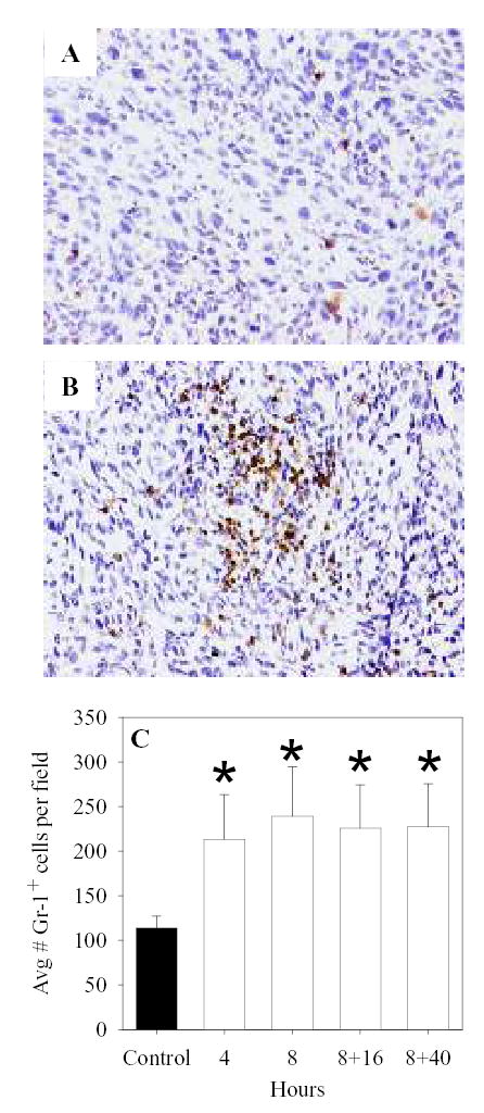
Increased numbers of Gr-1+ cells are observed in CT26 tumors of heated mice. After CT26 tumors were established s.c., BALB/c mice were heated (B, and white bars of C) or left as controls (A and black bar of C). At different times during and after and 8hr WBH, tumors were harvested, fixed, embedded in paraffin and then sectioned and stained for the granulocyte marker Gr-1. Average numbers of positively staining cells were determined per 40X field in three fields of sections made with three different tumors at each time point. *, p < 0.05 when compared to non-heated controls.