
Fig. 7.
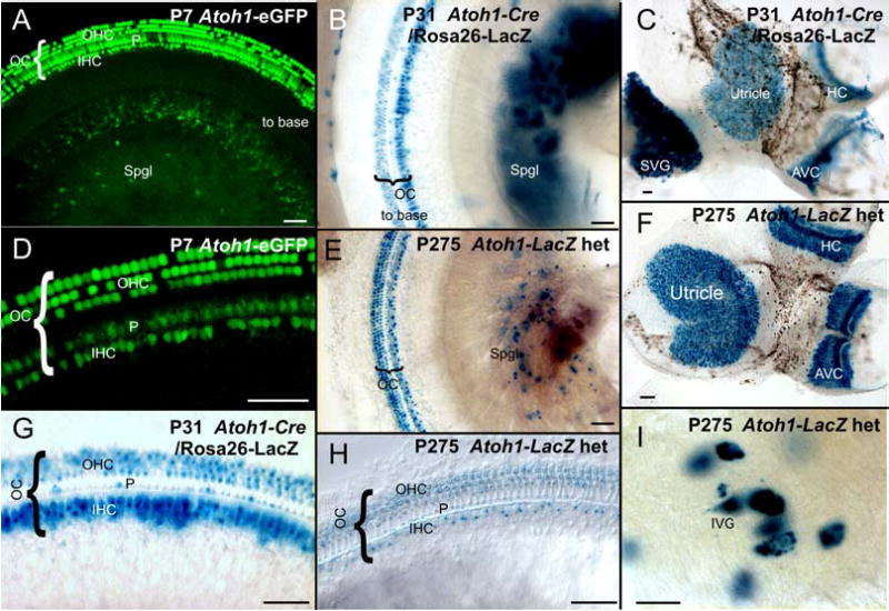
Expression of Atoh1 (AI) in hair cells and sensory neurons of juveniles and adults is shown as revealed with different techniques. With all these techniques, hair cells are most profoundly positive (D, C, F, G, H). However, additional cells in the cochlea are also positive for Atoh1 using either Atoh1-eGFP (A, D), Atoh1-Cre (B, C, G) or Atoh1-LacZ (H) and can be identified as pillar cells. Additional label is found in cells of the stria vascularis (near B). Note that Atoh1-LacZ staining in spiral or vestibular sensory neurons can be readily demonstrated in postnatal animals of all ages with any of these techniques (A, B, C, E, I). High power images show that the vestibular ganglion neurons (I) are approximately 50 μm in diameter and thus can not be confused with any other cell type in the vestibular ganglion. The similarities in detail of the sensory neuron expression obtained with all three techniques supports the idea that at least some vestibular and spiral neurons express Atoh1. However, the expression of β-galactosidase is particularly profound in spiral and vestibular neurons of Atoh1-Cre mice, suggesting that there may be a build up β-galactosidase over time, leading to a more obvious reaction in these large cells including a faint labeling in the nerve leading to the sensory epithelia. IVG, inferior vestibular ganglion; OC, Organ of Corti; S, saccule; Spgl, spiral ganglion; SVG, superior vestibular ganglion; U, utricle Bar indicates 50 μm in D, G, H, I and 100 μm in A, B, C, E, F.