
Fig. 9.
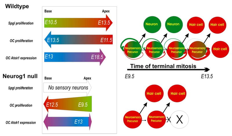
This summary diagram shows the spatiotemporal progression of sensory neuron and hair cell terminal mitosis, and Atoh1 expression in wildtype (top) and Neurog1 null mice (bottom). Our data and those of Ruben (1967) suggest that in wildtype the spiral ganglion terminal mitosis (Spgl) progresses from the base of the cochlea to the apex whereas the proliferation of hair cells of the organ of Corti progresses from apex to base with a broad overlap of terminal mitosis of both cell types in the apex around E11.5–12.5. The Neurog1 null ear has no spiral neurons, and shows a premature onset and cessation of terminal mitosis in hair cells in an apex to base gradient, along a time line comparable to sensory neurons in wildtype mice. Neurog1 null mice have fewer hair cells in a shortened and widened cochlea. Neurog1 null mice also show some alteration in the topology of Atoh1 expression. The right half of the scheme presents a hypothetical relationship between neurosensory precursors that give rise to both hair cells and sensory neurons. The model assumes that some neurosensory precursors will undergo a progressive up-regulation of Atoh1 in Neurog1 positive precursors until eventually they will turn these cells into hair cell producing precursors. It also assumes a mutual inhibition of Neurog1 and Atoh1 in the same precursor. According to this model, absence of NEUROG1 protein in Neurog1 null mutants causes premature commitment to hair cell fate and thus a terminal mitosis of hair cells at earlier developmental stages as well as truncation of the progenitor pool expansion, resulting in a shortened cochlea.
[modified after Ma et al., 1999; Gowan et al., 2001; Bertrand et al., 2002]