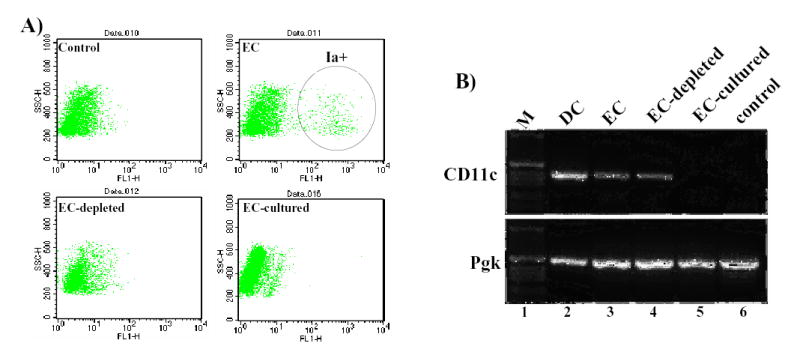
Figure 2. Fate of ex vivo transduced keratinocytes grafted to immunocompetent mice.

(A) Class II expression on mouse epidermal cells as assessed by flow cytometry. Analysis was performed on freshly isolated newborn epidermal cells after staining with isotype matched control antibody (control); anti mouse Ia antibody (EC); anti-mouse Ia antibody following treatment with anti-MHC classII magnetic beads (EC-depleted); and anti-mouse Ia antibody following 3 days of culture (EC-cultured). (B) RT-PCR analysis of APC contamination in epidermal cultures. Total RNA samples from epidermal dendritic cells islolated with Ia-specific immunobeads (DC), freshly isolated epidermal cells (EC), anti-Ia immunobead-depleted cells (EC-depleted), cells cultured for 7 days (EC-cultured) and immortalized mouse keratinocytes by RT-PCR using a set of primers specific for CD11c (upper panel) or phosphoglycerate kinase-1 (lower panel).
