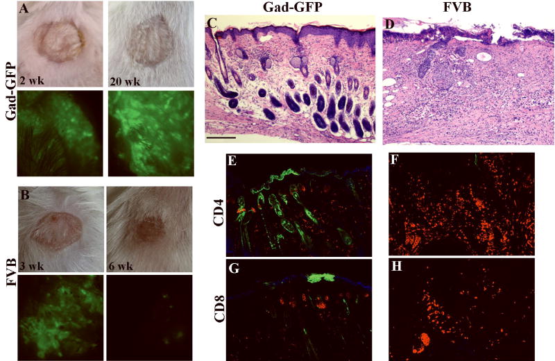
Figure 4. GFP expression in transduced cells grafted onto immunocompetent mice.
Cultured epidermal cells were transduced with LZRS-GFP at MOI of 2 and grafted onto FVB or GFP-tolerant FVB mice (Gad-GFP). Gross appearance of grafts and surface GFP expression at indicated time points are shown in Gad-GFP mice (A) and FVB mice (B). Surface GFP is lost in FVB mice at 6 weeks post-grafting (B). Hematoxylin/eosin histological sections (C-D) and immunostaining with anti-CD4 or anti-CD8 antibodies (red fluorescent staining) of grafted skin taken at 6 weeks post grafting from Gad-GFP (C, E, G) or FVB (D, F, H). GFP expressing cells appeared as green and were present in epidermis, sebaceous glands and hair follicles formed by the grafted cells in Gad-GFP mice (E and G). No GFP positive cells were present in the LZRS-GFP-transduced skin of FVB mice (F and H). Tissue sections were counterstained with DAPI (blue nuclear staining in J and L). Bar=100 μm for C-D, and 70 μm for E-H.

