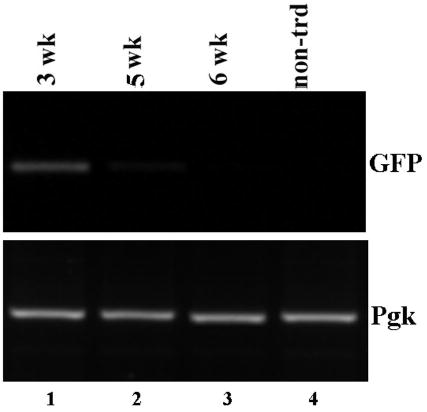
Figure 5. Loss of surface GFP correlates with the loss of transduced cells.

PCR analysis of genomic DNA isolated from skin grafts reconstituted from transduced keratinocytes. Skin tissues were harvested at 3, 5 and 6 weeks post-grafting (lanes 1–3), genomic DNA were isolated and analyzed by PCR using primers specific to GFP (top panel) or phosphoglycerate kinase (lower panels). DNA isolated from non-transduced skin (lane 4) was used as a control.
