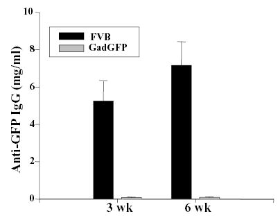
Figure 6. Immune activation in FVB mice grafted with transduced keratinocytes.

FVB or Gad-GFP mice were grafted with keratinocytes transduced with LZRS-GFP. Sera was collected at 3 or 6 weeks post-grafting and assessed for the presence of anti-GFP IgG by ELISA. The concentration of anti-GFP IgG is expressed based on the concentration of monoclonal anti-GFP antibody used as a standard in the ELISA. Error bar indicates standard deviation for each group (n = 10).
