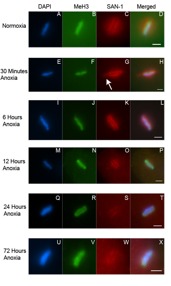
Figure 5.

SAN-1 localization to the kinetochore of metaphase blastomeres is reduced in embryos exposed to prolonged anoxia. Enlarged image of metaphase blastomeres of 4–8 cell embryos exposed to either normoxia (A-D), 30 minutes (E-H), 6 hours (I-L), 12 hours (M-P), 24 hours (Q-T), or 72 hours (U-X) of anoxia, collected, and stained with DAPI (A, E, I, M, Q, U), antibody MeH3, which recognizes methylated Histone H3 (B, F, J, N, R, V), and an antibody that recognizes SAN-1 (C, G, K, O, S, W). Merged image for each set is shown (D, H, L, P, T, X). White arrow points to the lateral flare detected in metaphase blastomeres of 4-cell embryos exposed to anoxia. For all images the scale bar equals 2 μm.
