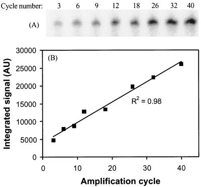
FIG. 5.
Linear amplification with 5′-32P-labeled primer 104r. (A) Gel electrophoresis and phosphorimaging detection of amplified fragments after 3 to 40 cycles. (B) Quantification by image analysis using local background subtraction shows a linear accumulation of amplification product with cycle number. AU, absorbance units.

