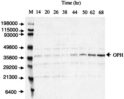
FIG. 4.
Western blot analysis of fermentation samples. Total cell pellets were diluted to approximately 0.2 g (dry cell weight)/liter and then boiled on SDS-PAGE cracking buffer and resolved on a 12% polyacrylamide gel. The proteins were transferred to a nitrocellulose membrane, and blotting was done with anti-OPH antibody. The figure shows the time course of OPH production. M, broad-range MW marker, with MWs indicated on the left.

