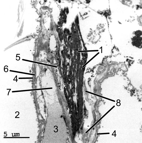
Figure 2.
EM morphology of the postmortem ELS lung (the same case as shown in Figure 1). The ELS are seen within the alveolar septum bordered by the basement membrane of the alveolar space on each side. The EM image dramatically demonstrates the in situ nature of the ELS: 1 = ELS; 2 = alveolar space; 3 = erythrocyte; 4 = basement membrane; 5 = endothelial cell nucleus; 6 = endothelial cell plasma membrane; 7 = capillary plasma; 8 = collagen fibers. (50,000×)

