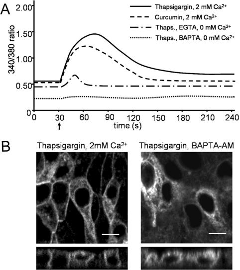
Figure 6.
Cytosolic calcium responses to treatment with SERCA inhibitors. (A) MDCK type 1 cells were seeded at 100,000 cells/cm2 in glass-bottom culture dishes and grown for 24 h. Cytosolic calcium levels were measured using FURA-2. Cells were incubated in HEPES/Tris buffer with the indicated levels of Ca2+, or EGTA with or without BAPTA (indicated), to allow the baseline to settle. Thirty seconds after the start of the measurement (indicated with an arrow), 1 μM thapsigargin or curcumin (indicated) was added to the cells followed by measurement of the 340/380-nm ratio. Each data series comprises the averaged data of 25 cells that were measured simultaneously. The figures show data of a representative experiment (n = 3). (B) Confluent MDCK-V2R-V206D cells were incubated for 2 h in HEPES/Tris buffer with 1 μM thapsigargin, supplemented with 2 mM Ca2+ (left), or 5 mM BAPTA-AM and without Ca2+ (right). Subsequently, cells were fixed and analyzed by confocal microscopy. Bar, 10 μM.

