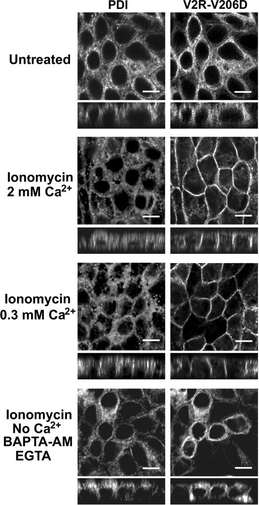
Figure 7.
Effect of ionomycin and calcium on the localization of V2R-V206D. Confluent cell layers of MDCK cells stably expressing GFP-tagged V2R-V206D were incubated for 2 h in HEPES/Tris buffer with 2 mM Ca2+, 2 mM Ca2+, and 1 μM ionomycin, or 300 μM Ca2+ and 1 μM ionomycin, or in buffer without Ca2+ in the presence of 1 μM ionomycin, 5 mM BAPTA-AM, and 1 mM EGTA (indicated). Subsequently, the cells were fixed, immunocytochemically stained, and analyzed by CLSM as described in the legend of Figure 2. Bar, 10 μm.

