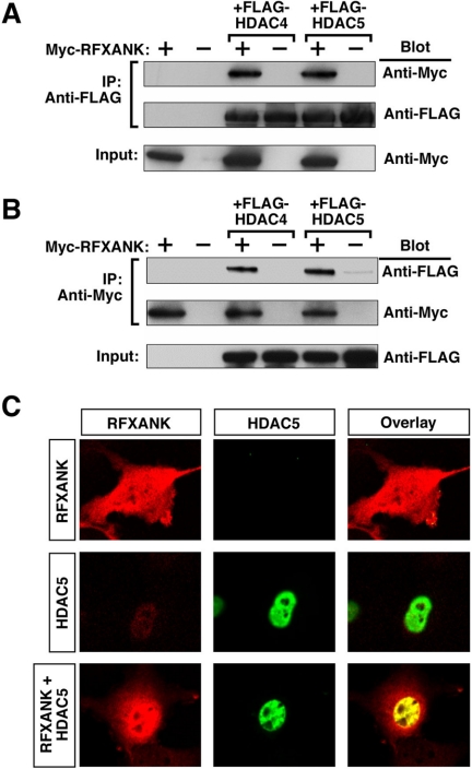
Figure 5.
Interaction of RFXANK with HDAC4 and HDAC5 in mammalian cells. (A) 293T cells were cotransfected with expression vectors encoding FLAG-tagged HDAC5 and Myc-RFXANK. Cell extracts were immunoprecipitated (IP) with anti-FLAG antibodies and proteins in the immune complexes were analyzed by Western blotting with the indicated antibodies (Blot). Crude lysates were analyzed by Western blotting to control for variability in protein expression (Input). (B) 293T cells were transfected and protein lysates prepared as described in A. IP was performed with anti-Myc antibody and proteins in immune complexes were analyzed by Western blotting as described above. (C) Colocalization of HDAC5 with RFXANK. COS cells were transfected with expression vectors encoding either FLAG-tagged HDAC5 or Myc-tagged RFXANK. Some cells were cotransfected with these vectors (RFXANK + HDAC5). The subcellular localization of RFXANK and HDAC5 was determined by indirect immunofluorescence. Proteins were visualized with a confocal microscope and digital images captured (red, RFXANK; green, HDAC5; yellow, colocalized RFXANK and HDAC5).

