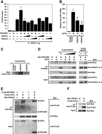
Figure 7.
HDAC4 and HDAC5 repress CIITA-inducible HLA-DRA promoter activity. (A) COS cells were transfected with a luciferase reporter controlled by the HLA-DRA promoter in the absence or presence of vectors for RFXANK (100 ng), CIITA (5 ng), or HDAC4 or HDAC5 (10 or 100 ng), as indicated. Cells were harvested for luciferase assay 48 h after transfection. (B) HeLa cells were transfected with expression plasmid encoding CIITA (10 ng) in the absence or presence of vectors for HDAC4 or HDAC5 (100 ng). Forty-eight hours later, RNA was prepared from the cells and HLA-DRA mRNA transcripts were detected by real-time RT-PCR. Values were normalized to those for 18S rRNA. (C) Hela cells were transfected as described in B. Protein lysates were analyzed by immunoblotting with anti-FLAG antibody to assess effects of HDAC overexpression on FLAG-CIITA levels. (D) 293T cells were transfected with the indicated combinations of expression vectors encoding CIITA, Myc-tagged RFXANK and FLAG-tagged HDAC4 or HDAC5. Cell extracts were immunoprecipitated (IP) with anti-Myc antibody and proteins in immune complexes were analyzed by Western blotting (Blot) with the indicated antibodies. Crude lysates were analyzed by Western blotting to control for variability in protein expression (Input). (E) 293T cells were transfected with expression vectors encoding FLAG-CIITA, FLAG-RFXANK, or myc-HDAC4 (1 μg each), as indicated. Cell extracts were immunoprecipitated with anti-Myc antibody and proteins were analyzed by immunoblotting with anti-FLAG antibody. (F) 293T cells were transfected with expression vectors encoding either FLAG-CIITA or Myc-HDAC4 (1 μg each), as indicated. Cell extracts were immunoprecipitated with anti-FLAG antibody and proteins were analyzed by immunoblotting with anti-Myc antibody.

