Figure 8.
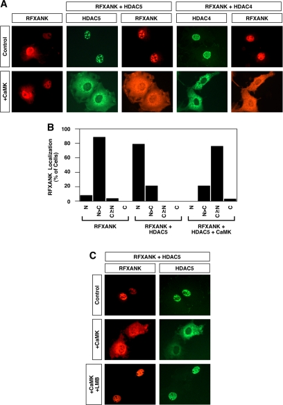
Nuclear export of class II HDAC/RFXANK complexes. (A) COS cells were transfected with an expression vector encoding Myc-tagged RFXANK in the absence or presence of vectors for FLAG-tagged HDAC5 or HDAC4. As indicated, some cells also received a plasmid encoding constitutively active CaMKI. The subcellular localization of RFXANK and HDACs was determined by indirect immunofluorescence (red, RFXANK; green, HDAC4 or HDAC5). (B) Effects of HDAC5 and CaMKI overexpression on RFXANK subcellular distribution were quantified by microscopic examination of greater than 100 cells per condition. N, exclusive staining of RFXANK in the nucleus; N>C, nuclear RFXANK staining greater than cytoplasmic staining; C≥N, cytoplasmic RFXANK staining greater than or equal to nuclear staining; C, exclusive staining of RFXANK in the cytoplasm. (C) COS cells were cotransfected with expression vectors for RFXANK and HDAC5 in the absence or presence of a plasmid encoding activated CaMKI. Forty-eight hours after transfection, cells were exposed to leptomycin B (LMB; 10 nM) for 2 h. The subcellular localization of RFXANK and HDAC5 was determined by indirect immunofluorescence.

