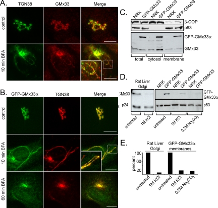
Figure 1.
Endogenous GMx33 and GFP-GMx33α are peripherally localized to the TGN. (A) NRK cells were left untreated (top panels) and treated with 5 μg/ml brefeldin A for 10 min (bottom panels), fixed, and stained with antibodies specific for either TGN38 or GMx33. Our GMx33-specific antibody recognizes both α and β forms of GMx33. Primary antibodies were detected with either anti-mouse or anti-rabbit secondary antibodies conjugated to either Alexa Fluor 488 or Alexa Fluor 594. The inset is an enlarged image from the area indicated by the dotted box. (B) NRK cells stably expressing GFP-GMx33α were left untreated (top panel) or treated with brefeldin A for 10 or 60 min (middle and bottom panels). Fixed cells were stained for TGN38. The inset is an enlarged image from the area indicated by the dotted box. (C) NRK cells stably expressing GFP-GMx33α were lysed in detergent (total) or used to prepare membrane and cytosol fractions as described in Materials and Methods. Protein (50 μg per lane) was analyzed by immunoblot with antibodies specific for the indicated proteins. (D) Fifteen micrograms of rat liver Golgi fraction were left untreated or stripped with 1 M KCl for 1 h on ice and washed (left panel). One hundred micrograms of total membrane fractions from wild-type NRK cells or those expressing GFP-GMx33α (right panel) were left untreated or stripped with 1 M KCl as above or a high pH wash, 0.2 M sodium carbonate for 15 min on ice. Half of each sample was subjected to SDS-PAGE and analyzed by immunoblot with antibodies specific for the indicated proteins. p24 and p63 proteins were followed as loading controls. (E) Densitometry was performed on the immunoblots in D using the NIH Image J software. The amount of p24 or p63 signal in each lane was used to normalize slight differences in loading. Bars, 10 μm.

