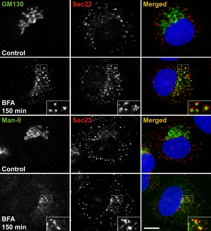
Figure 2.
Mannosidase II accumulates at ER exit sites at prolonged incubation with BFA. NRK cells were untreated (control) or treated with 5 μg/ml BFA for 150 min. Cells were fixed and processed for immunolabeling with antibodies against GM130 and Man-II (left), Sec23 (middle), and merged (right). Alexa Fluor-488 and Alexa Fluor-594 were used as fluorescent secondary probes. A hundred percent of the cells positive for GM130 (top two panels) and 30% of the cells positive for Man-II (bottom two panels) showed accumulation of these proteins in puncta after 150 min of BFA treatment. All GM130- or Man-II-positive puncta are near and/or at ER exit sites, defined by colocalization with Sec23, a component of the COPII coat (middle and merged images on right). Boxed regions are shown at higher magnification in the lower right corner. Bar, 10 μm.

