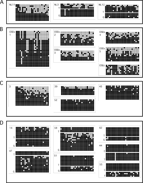
Figure 4.
Analysis of M6P/IGF2R region 2 methylation status by cloning and sequencing individual alleles from HTBM-treated DNA. Each row of squares corresponds to one individual clone; columns represent each cytosine in CpG context within the sequenced region. Filled box, methylated; unfilled, unmethylated. (A) Normal peripheral blood lymphocytes (NL) from women without evident malignancy. NL3 and NL12 were heterozygous for a T/G SNP within the sequenced region allowing the individual parental alleles to be identified. (B) Sequenced clones from normal ovarian surface epithelium (OSE). OSEs denotes samples that were obtained by gently scraping the surface epithelium from the ovary during surgical oophorectomy; OSEc are normal OSE cells that underwent several passages in tissue culture prior to DNA extraction and analysis. (C) Methylation status of alleles cloned from ovarian malignancies that exhibited differential methylation (3) and hypermethylation (39, 52, 43) by MS-PCR. (D) Methylation status of cloned alleles from ovarian malignancies heterozygous for one of two polymorphisms (a T/G or a C/T SNP) present within the sequenced region, allowing for discrimination between parental alleles; all of these specimens exhibited hypermethylation by MS-PCR.

