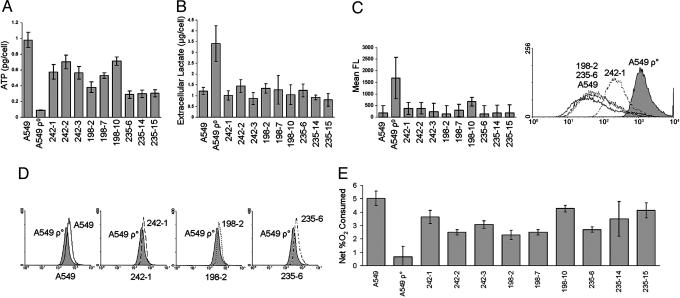
Fig. 4.
Recovery of mitochondrial function in rescued clones. A549 cells, A549 ρ° cells, and rescued clones were analyzed for mitochondrial function by several assays. (A) Without exception, the rescued clones had significantly increased intracellular ATP compared with A549 ρ° cells. (B) Extracellular lactate was significantly reduced in the rescued clones when normalized for cell number, reaching levels comparable to parental A549 cells. (C) There was a decrease in reactive oxygen species compared with A549 ρ° cells in all of the rescued clones. Values (Left) are relative levels of mean fluorescence (FL) derived from the histogram shown (Right). (D) There was an increase in mitochondrial membrane potential in all rescued clones compared with A549 ρ° cells (242-1, 198-2, and 235-6). (E) Rescued clones consumed almost as much O2 as the parental A549 cells, indicating aerobic respiration. Error bars represent one standard deviation from the mean of three individual cultures (A–D) or three measurements of a single culture (E). Two-tailed Student t tests for A, B, and E indicated P values <0.05.

