FIG. 2.
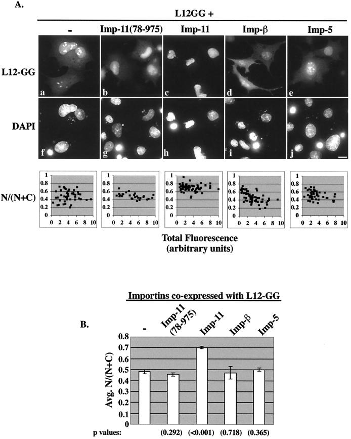
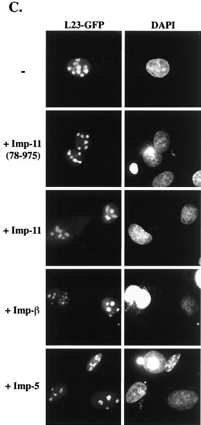
Importin 11 selectively increases the nuclear accumulation of rpL12. (A) Transiently transfected BHK cells ectopically expressing L12-GG alone or with the indicated HA-tagged importins were fixed, permeabilized, DAPI stained, and analyzed by fluorescence microscopy. Representative cells from each sample are shown. Panels a to e show the GFP fluorescence (L12-GG), and panels f to j show the DNA staining (DAPI). Bar, 10 μm. Corresponding graphs show fractional nuclear GFP fluorescence for a range of expression levels of L12-GG, in the presence or absence of the coexpressed importins. GFP images were captured such that no pixels were saturated and quantitated to obtain total fluorescence (N + C) and nuclear fluorescence (N) for each cell. The data were compiled from 25 to 50 cells for each sample. The L12-GG images shown were all captured with identical camera settings and exposure times and adjusted to the exact same settings using Photoshop software. As a result of these postquantitative adjustments, some pixels appear to be saturated, especially in the nucleoli. (B) Average N/(N + C) calculated from the data presented in the above scatter graphs. Standard error bars are indicated for each set of data, and statistical significance was determined by comparing the N/(N + C) values obtained for L12GG alone to those from each of the other conditions by using Student's t test. P values are indicated in parentheses below each bar of the graph. (C) BHK cells ectopically expressing a GFP fusion of ribosomal protein L23a (L23-GFP) alone or with the indicated HA-tagged importins. Cells were processed as described for panel A. The left panels show the GFP fluorescence (L23-GFP), and the right panels show the DNA staining (DAPI).


