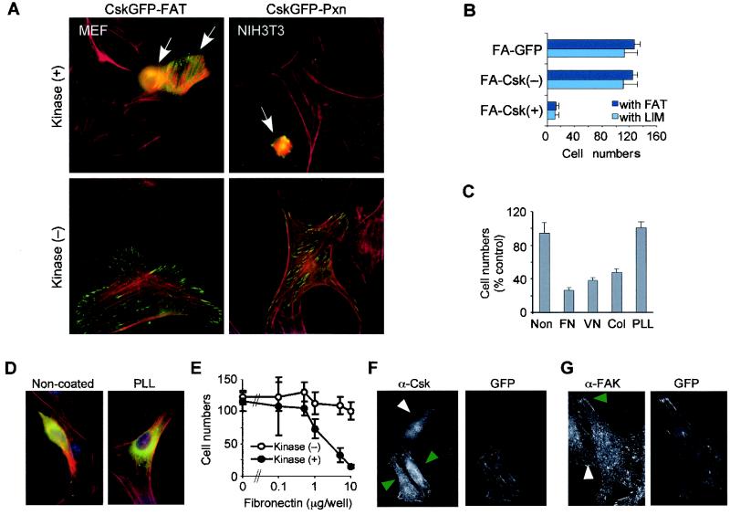
FIG. 2.
Loss of integrin-mediated adhesion caused by FA-Csk. (A) The morphologies of wild-type MEFs and NIH 3T3 cells expressing kinase-active or -inactive FA-Csk constructs were determined after transient expression of the transgene. Only one example of each construct with each cell type is shown. Phalloidin staining is shown in red. Green indicates GFP fluorescence in the transfected cell (arrows). (B) The numbers of GFP-positive NIH 3T3 cells remaining on fibronectin-coated coverslips were determined in 30 random fields under a 25× objective and expressed as percentages (± standard deviations [SD] in a triplicate experiment) of the control group electroporated with GFP alone. (C) The numbers of GFP-positive NIH 3T3 cells remaining attached were determined on an ECM protein or PLL (PLL). Cells were transfected by lipofection with CskGFP-FAT. FN, fibronectin; VN, vitronectin; Col, denatured collagen (gelatin). The values indicate the numbers of GFP-positive cells relative to those of the control group transfected with the kinase-inactive variant. (D) Morphology of NIH 3T3 cells expressing the kinase-active version of CskGFP-FAT on PLL-coated glass coverslips. Green and red indicate GFP fluorescence and F-actin, respectively. (E) NIH 3T3 cells were plated on glass coverslips coated with increasing concentrations of fibronectin prior to transient transfection with 0.1 μg of CskGFP-FAT construct per well by lipofection. The y axis shows the total number (mean ± SD in a triplicate experiment) of GFP-positive cells remaining on the tissue culture surface in each group. (F) NIH 3T3 cells were stained with anti-Csk antibody (α-Csk) after electroporation with kinase-inactive CskGFP-FAT in order to determine expression levels of the transgene compared to endogenous Csk. Green and white arrowheads indicate cells transfected and nontransfected, respectively. Levels of proteins identified by anti-Csk antibody were measured as described in Materials and Methods. (G) NIH 3T3 cells were plated on fibronectin-coated coverslips and stained with an anti-Fak antibody (α-Fak) after electroporation with kinase-negative CskGFP-FAT. This antibody detects endogenous Fak but not the fusion protein, as it recognizes the N terminus of Fak. Green or white arrowheads indicate endogenous Fak at FA structures in a transfected cell or in a nontransfected cell, respectively. In all experiments shown (panels A to G), cells were cultured in DMEM containing 10% calf serum for 18 h after transfection before fixation. All experiments were repeated at least three times.

