FIG. 1.
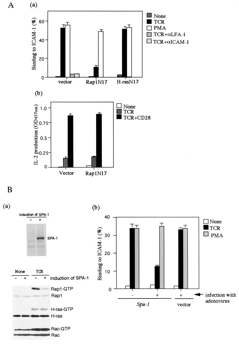
TCR-induced adhesion of Jurkat cells to ICAM-1 is inhibited by Rap1N17 or SPA-1. (Aa) Adhesion of Jurkat cells transfected with vector alone, Rap1N17, and H-rasN17. Jurkat cells were stimulated with or without 1 μg of OKT3 (TCR) or 10 ng of PMA per ml for 30 min at 37°C in ICAM-1-coated plates, as described in Materials and Methods. The level of adhesion to BSA was <1%. Average and standard errors of triplicate experiments are shown. (Ab) IL-2 production upon stimulation with TCR-CD28 antibody cross-linking. Rap1N17 or pcDNA3 (vector) stable transfectants were stimulated by cross-linking of the TCR complex with OKT3 (TCR) or OKT3 and CD28.2 (TCR+CD28) for 16 h, and the supernatants were harvested for IL-2 measurement. An optical density at 437 nm of 1 was equal to 0.15 ng of recombinant mouse IL-2/ml. (Ba) The upper panel shows the induction of SPA-1 detected with anti-flag antibody. SPA-1 was uninduced (−) or induced (+) by infection with adenovirus expressing cre recombinase for 2 days. The lower panel shows the inhibition of TCR-mediated Rap1 activation by the induction of SPA-1 expression. Jurkat cells uninduced (−) or induced (+) to express SPA-1 were stimulated with OKT3 (TCR) for 10 min and lysed, and GTP-bound Rap1, H-ras, and Rac were detected with pull-down assays by using immobilized GST fusion proteins of RalGDS-RBD, Raf-RBD, and PAK-CD. Western blots of total cell lysates are also shown. (Bb) Adhesion of Jurkat cells uninduced (−) or induced (+) to express SPA-1 with OKT3 (TCR) or PMA. Jurkat cells transfected with Spa-1 or empty vector were infected with adenovirus carrying cre recombinase. Infected cells were stimulated with or without 1 μg of OKT3 (TCR) or 10 ng of PMA per ml for 30 min at 37°C in ICAM-1-coated plates, as described in Materials and Methods. Average and standard errors of triplicate experiments are shown. (Ca) Decrease in TCR-dependent Rap1 activation by costimulation with antibody cross-linking of CD28. Jurkat cells were stimulated with OKT3 in the absence (TCR) or presence of CD28.2 (TCR/CD28) for 10 min and lysed, and GTP-bound Rap1, H-ras, and Rac were measured as described in Fig. 1Ba. (Cb) TCR-induced adhesion of Jurkat cells was reduced by costimulation with CD28. Jurkat cells were unstimulated or stimulated with OKT3 (TCR), CD28.2 (CD28), or TS2/18 (CD2) alone or in combination for 30 min at 37°C in ICAM-1-coated plates. Average and standard errors of triplicate experiments are shown.

