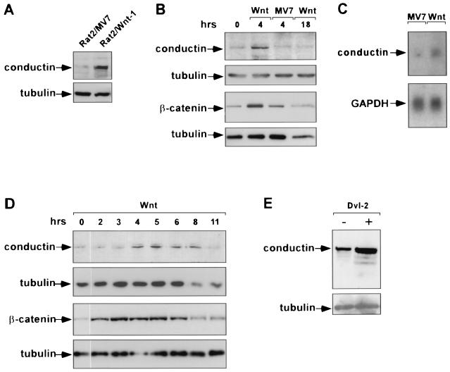
FIG. 5.
Upregulation of conductin by Wnt signaling. (A) Expression of conductin in Rat2/Wnt-1 cells and Rat2/MV7 control cells demonstrated by Western blotting using the C/G7 antibody. Note increased levels of conductin in the Wnt-1-expressing cells. (B) Upregulation of conductin in MDA MB 231 breast carcinoma cells by Wnt-1. Cells were incubated with media conditioned by Rat2/Wnt-1 (Wnt) or Rat2/MV7 cells (MV7) for 4 and 18 h as indicated above the lanes. Conductin levels were determined by Western blotting as described for panel A. Western blotting for β-catenin was performed on cytosolic extracts prepared from parallel cultures. Note that both conductin and β-catenin levels increase after 4 h of stimulation by Wnt-1-conditioned medium and decrease to baseline after 18 h. Control conditioned medium had no effect. (C) Increase in conductin mRNA after treatment of MDA MB 231 cells with Wnt-1-conditioned medium for 4 h as determined by Northern blotting. GAPDH, glyceraldehyde-3-phosphate dehydrogenase. (D) Time course of upregulation of conductin and β-catenin by Wnt-1 in MDA MB 231 cells as determined by Western blotting. In this experiment, both conductin and β-catenin were detected from the same cytosolic extracts. Note that β-catenin peaks between 2 and 6 h while conductin peaks between 4 and 8 h after stimulation with Wnt-1. Tubulin was probed to demonstrate protein loading. (E) Upregulation of conductin after transient expression of dishevelled-2 (Dvl-2) in Neuro2A cells. Conductin and tubulin were detected by Western blotting.

