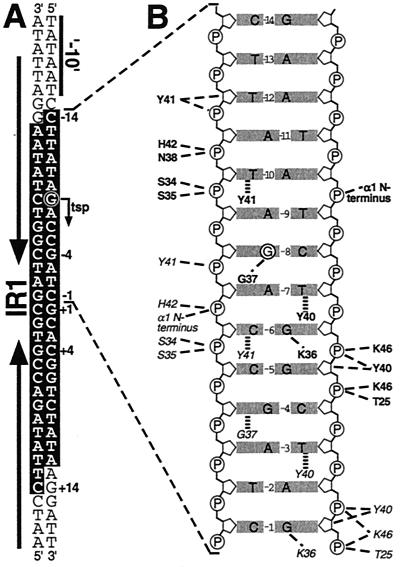
FIG. 12.
DNA contacts formed by QacR to an IR1 half-site. (A) Sequence of the qacA −10 promoter region and the downstream IR1 operator, with the bases that QacR protects from DNase I digestion highlighted (white) against a black background, the qacA transcription start point (tsp) circled, and the location of IR1 indicated by bold arrows that flank the central 6-bp IR1 spacer region (47). (B) The DNA contacts made by a pair of operator-bound QacR dimers to a single IR1 half-site. The bp −1 to −14, which constitute the depicted operator half-site, are shown as a mirror image of A. The QacR residues in bold that contact the DNA in this half-site are from the distal subunit of dimer 2, whereas those in italics are from the proximal subunit of dimer 1 (Fig. 11, purple and green polypeptides, respectively). The contacts made to the bases and DNA backbone in the operator half-site by these amino acids are shown as thin dashed lines for hydrogen bonds and thick broken lines for van der Waals interactions. Also indicated is a DNA contact made by the N terminus of α1. (Panel B reprinted with permission from reference 197.)

