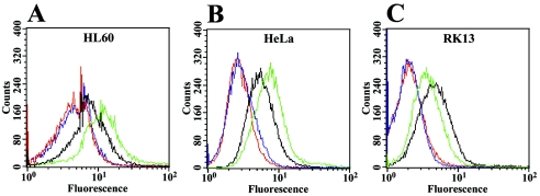
FIG. 3.
Effect of PMA on RPTPβ protein in HL-60 (A), HeLa (B), or RK13 (C) cells. Cells were incubated for 24 h with (green lines) or without (black lines) 20 nM PMA, and RPTPβ expression was quantified by indirect immunofluorescence and flow cytometry using anti-RPTPβ monoclonal antibodies. An irrelevant mouse IgG was used as a control on cells with (blue lines) or without (red lines) 20 nM PMA. Data are representative of three separate experiments.

