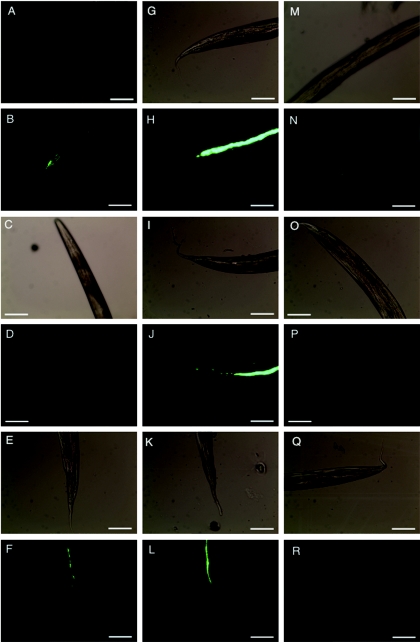
FIG.4.
EPEC persistence within the C. elegans gut. Synchronized L4 nematodes were subjected to infection with GFP-labeled, pGEN91-containing strain E2348/69 (A to F), TB135A (G to L), or MG1655 (M to R) on NGM agar supplemented with the appropriate antibiotics. At 24 hours postinfection, nematodes were rinsed gently with M9 buffer and then transferred to NGM agar containing lawns of the feeding strain OP50 without antibiotic selection. Nematodes were prepared for light and fluorescence microscopy as described in Materials and Methods 0 (A, B, G, H, M, N), 24 (C, D, I, J, O, P), and 48 (E, F, K, L, Q, R) hours after transfer to coculture with strain OP50. Light microscopy images (A, C, E, G, I, K, M O, and Q) appear adjacent to corresponding fluorescence microscopy images (B, D, F, H, J, L, N, P, and R). Representative images are presented. Magnification, ×200. Bars, 100 μm.

