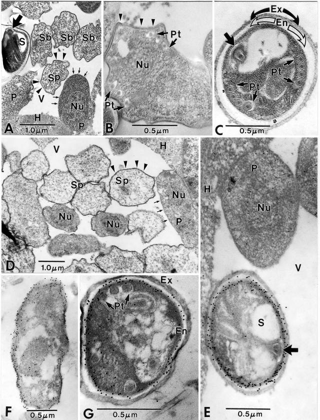
FIG.6.
Immunoelectron microscopy of Encephalitozoon cuniculi using anti-ECU01_1270. (A to C) Control sections of Encephalitozoon cuniculi-infected rabbit kidney cells. (A) Low-magnification image of a portion of host cell cytoplasm (H) containing a parasitophorous vacuole (V) with proliferative parasite cells (P) abutted to the edge of the host-parasite interface. These cells have a simple cytoplasm, are uninucleate (Nu), and have a typical “thin” plasmalemmal surface (short arrows). The sporonts (Sp) and sporoblasts (Sb) have a “thick” dense surface coat (arrowheads) and a more complex and dense cytoplasm. The spore (S) contains a polar tube, and the anterior anchoring disk (broad arrow) is present. (B) Sporoblast containing portions of the developing polar tube (Pt), a single nucleus (Nu), and a “thick” surface coat (arrowheads). Note the irregular shape of the cell and the more complex cytoplasm. (C) Spore demonstrating groups of four cross sections of the polar tube (Pt) and its anterior anchoring disk (broad arrow). The sporoplasm is enclosed by a wide electron-lucent endospore (En), which is in turn covered by a dense, thick exospore (Ex). Note the absence of gold particles in all three images (A, B, and C). (D to G) Sections of Encephalitozoon cuniculi-infected rabbit kidney cells incubated with anti- ECU01_1270. (D) Portion of a parasitophorous vacuole (V) in the host cell cytoplasm (H) with proliferative parasite cells (P) abutted to the edge of the host-parasite interface. These cells are uninucleate (Nu) and have a typical “thin” plasmalemmal surface (short arrows). The sporonts (Sp) have a “thick” dense surface coat (arrowheads) and a more complex and dense cytoplasm. Colloidal gold particles (12 nm) are dispersed throughout the cytoplasm of the proliferative cells, while the sporogonic cells tend to have more gold localized near their plasmalemmal surfaces. There are a few gold particles at the host cell-parasite interface, but the vacuolar space (V) is free of gold. (E) High-magnification image of a portion of the host cell cytoplasm (H) and a parasitophorous vacuole (V) containing a proliferative parasite cell (P) abutted to the edge of the host-parasite interface. This uninucleate (Nu) cell has several gold particles in its cytoplasm and in the vicinity of the nucleus. The spore (S) with an anchoring disk (broad arrow) next to the proliferative cell has extensive quantities of gold distributed almost exclusively along the periphery of the plasmalemma-endospore interface. The host cell cytoplasm has a few scattered gold particles at the interface with the vacuolar space. (F) Sporont with extensive gold particles distributed along the periphery of the cell and abutting the plasmalemma and the overlying “thick” surface coat that is present during the transition to sporogony. The cytoplasm still has some diffuse gold particles in it. (G) Typical late sporoblast/early spore containing several cross sections of the polar tube and a dense sporoplasm enclosed by a well-defined plasmalemma abutted to a wide electron-lucent endospore (En) that is in turn covered by the dense exospore (Ex). A few gold particles are present in the sporoplasm, but the vast majority of them are distributed at the endospore-plasmalemma interface.

