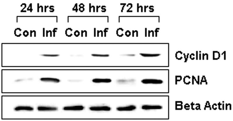
FIG. 1.

Representative immunoblot demonstrating the expression of cyclin D1 and PCNA in infected smooth muscle cells. Lysates of smooth muscle cells were probed with antibodies directed against cyclin D1 and PCNA. Note that infected cells (Inf), at 24 h, 48 h, and 72 h postinfection, exhibited increased cyclin D1 and PCNA expression compared to control cells (Con). β-Actin was used as a loading marker.
