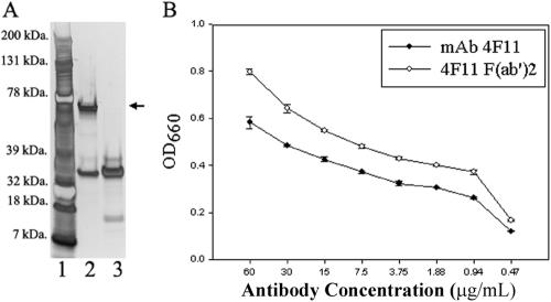
FIG. 1.
(A) SDS-PAGE analysis of intact MAb 4F11(G1) and 4F11(G1) F(ab′)2 fragments. Lane 1, molecular mass markers; lane 2, intact MAb 4F11(G1); lane 3, 4F11(G1) F(ab′)2 fragments. One microgram of total protein was added to each well. The arrow indicates the IgG heavy chain. (B) ELISA using soluble P. carinii antigens from infected mouse lung homogenates as target and MAb 4F11(G1) and 4F11(G1) F(ab′)2 as primary antibodies. Results represent means ± standard deviations of duplicate experiments with each condition tested in triplicate.

