Fig. 1.
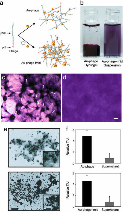
Concept and biological/structural characterization of Au–phage and Au–phage–imid networks. (a) Strategy for Au assembly onto phage nanoparticles. Imid and the yellow spheres [Au nanoparticles (not drawn to scale); the Au particles have a diameter of 44 ± 9 nm, and the pVIII capsid peptide has a thickness of ≈6 nm]. (b) Vials of nanoparticle solutions: Au–phage hydrogel (Left) and suspension of purified Au–phage–imid (Right; suspended from hydrogels precursor). (c and d) Hydrogel formed with RGD-4C-displaying phage. (Scale bar, 20 μm.) (c) C17.2 murine neural stem cells cultured within hydrogel for 24 h. Cell accumulation followed by cell-induced network displacement is shown (arrows point to cells within the network). (d) Control hydrogel (no cells). (e) TEM of purified networks: Au–phage (Upper) and Au–phage–imid (Lower). (Scale bar, 500 nm; inset scale bar, 100 nm.) (f) Bacterial infection with purified Au–phage (Upper) and Au–phage–imid (Lower) networks; TU are shown for purified and functional Au–phage and Au–phage–imid solution and for unbound phage present in the supernatant from centrifuged network solutions.

