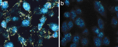
Fig. 6.
Dark-field images (real color) of cell-bound Au–phage networks using light from a microscope mercury lamp. Confluent KS1767 cells incubated with phage preparations (input of 1.0 × 10⁷ TU): Au–RGD-4C (gold color) (a) and Au–fd-tet (control insertless phage) networks (b). The blue color shows residual fluorescence from DAPI-stained cell nuclei.

