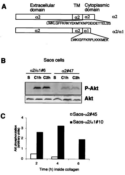
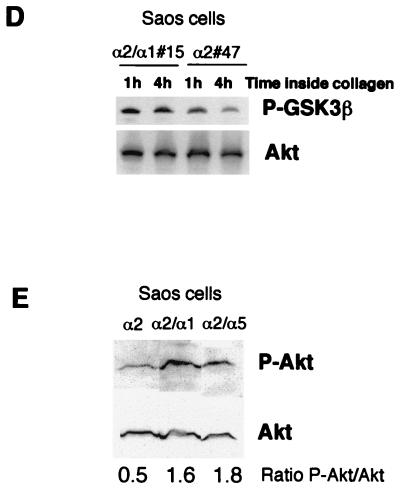
FIG. 2.
Collagen-induced dephosphorylation of Akt is regulated by α2β1 integrin in human osteosarcoma Saos cells. (A) cDNA constructs used for expression of integrin α2 and α2/α1 (chimera) subunits in Saos cells, which lack endogenous α2β1 integrin. TM, transmembrane domain. (B to E) Saos-α2, Saos-α2/α1, and Saos-α2/α5 cell clones were serum starved, detached, and cultured inside 3D collagen gel (C, collagen) for the indicated times or left in suspension (S) for 1 h. For panel E cells were cultured inside collagen for 2 h. Western blot analysis with antibodies that recognize Akt (B and E) and GSK3β (D) when they are phosphorylated (P-Akt and P-GSK3β, respectively) is shown. (B, D, and E) Immunoblotting with an antibody that recognizes all forms of Akt was performed for reference. (C) A separate experiment with longer incubation times inside collagen was performed with different Saos cell clones. Densitometric quantitation of phosphorylated Akt levels at different time points relative to loading is shown. If the experiment was performed with several cell clones, the clone numbers are indicated (e.g., #6).


