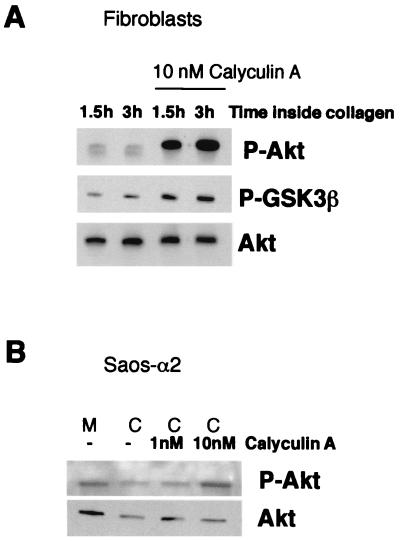
FIG. 3.
Regulation of Akt phosphorylation involves serine/threonine phosphatase activity. (A) Western blot analysis with an antibody that recognizes Akt or GSK3β when they are phosphorylated (P-Akt and P-GSK3β, respectively). Fibroblasts were deprived of serum for 16 h and seeded inside 3D collagen gel in the presence or absence of serine/threonine phosphatase inhibitor calyculin A (10 nM). (B) Western blot analysis with an antibody that recognizes phosphorylated Akt. Saos-α2 cells were deprived of serum for 16 h and then harvested from the cell culture dish (M, monolayer) and seeded inside 3D collagen (C) gel for 2 h. Cells were cultured in the presence or absence of serine/threonine phosphatase inhibitor calyculin A (1 and 10 nM). Immunoblotting with an antibody that recognizes all forms of Akt was performed for reference (A and B).

