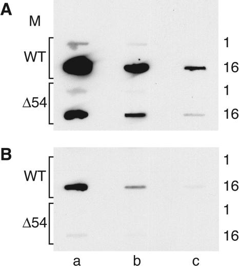
FIG. 11.
Analysis of DNA replication in infected PK(15) cells. PK(15) cells were mock infected (M) or infected with WT or vJSΔ54 (Δ54) at a MOI of 10 (A) or 0.01 (B). Total infected cell DNAs were harvested at 1 or 16 hpi and denatured, and serial 10-fold dilutions were slot-blotted onto a nylon membrane. Lane a represents 1.5 × 10⁵ cell equivalents of total cell DNA; lanes b and c contain 10−1 and 10−2 dilutions, respectively. Viral DNA was detected by hybridization with the biotinylated DNA probe, P1 (Fig. 1). Biotinylated DNA was visualized using a NEB Phototope kit as described in Materials and Methods.

