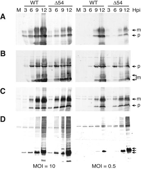
FIG. 6.
Accumulation of late proteins after infection with vJSΔ54. PK(15) cells were either mock infected (M) or infected at a MOI of 10 or 0.5. At 3, 6, 9, and 12 hpi, whole-cell extracts were prepared and subjected to analysis by SDS-PAGE. Protein accumulation was determined by Western blot analysis using antibodies specific for gC (A), gB (B), gE (C), or Us9 (D). The different glycoprotein isoforms (m, mature; p, precursor) are indicated by the arrows.

