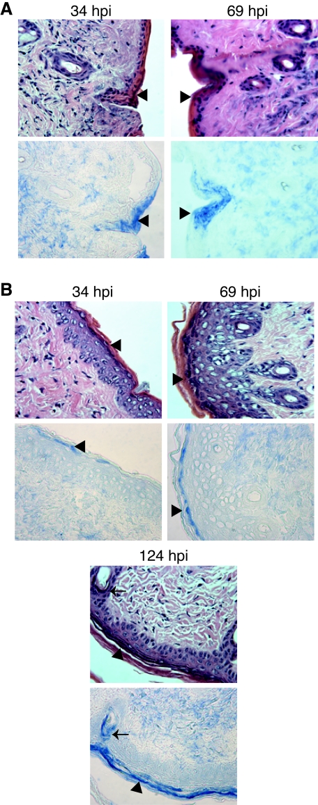
FIG. 8.
Distribution of PRV antigens in infected mouse skin tissue. Mice were infected with WT (A) or vJSΔ54 (B) virus as described in the legend of Fig. 7. At 34, 69, or 124 hpi, mice were euthanized, and skin adjacent to the inoculation site was dissected and fixed in 10% formalin. The tissues were embedded in paraffin, and 1-μm serial sections were collected. The sections were deparaffinized and subjected to immunohistochemical analysis. One serial section (top row) was counterstained with H&E, while the adjacent section (bottom row) was stained with the rabbit polyclonal antibody Rb133 (7), specific for whole PRV. Anti-PRV antibody was visualized with secondary antibodies conjugated to alkaline phosphatase and reacted to a blue substrate. Images were obtained with a 40× objective using a Leitz Laborlux microscope. The arrow identifies a hair follicle stained for PRV antigens, whereas the arrowheads identify epidermal cells containing PRV.

