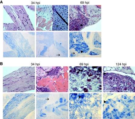
FIG. 9.
Distribution of PRV antigens in infected mouse DRGs. Mice were infected with WT (A) or vJSΔ54 (B) virus as described in the legend of Fig. 7. At 34, 69, or 124 hpi, mice were euthanized, and DRGs innervating the inoculation site dermatomes were dissected. The tissues were fixed, embedded, sectioned, and subjected to H&E staining or immunohistochemical analysis as described in the legend of Fig. 8. The leftmost panels represent nerve fiber associated with the dissected DRGs harvested at 34 hpi. The arrows indicate neurons that do not contain PRV antigens, while the arrowheads identify those that do contain PRV.

